
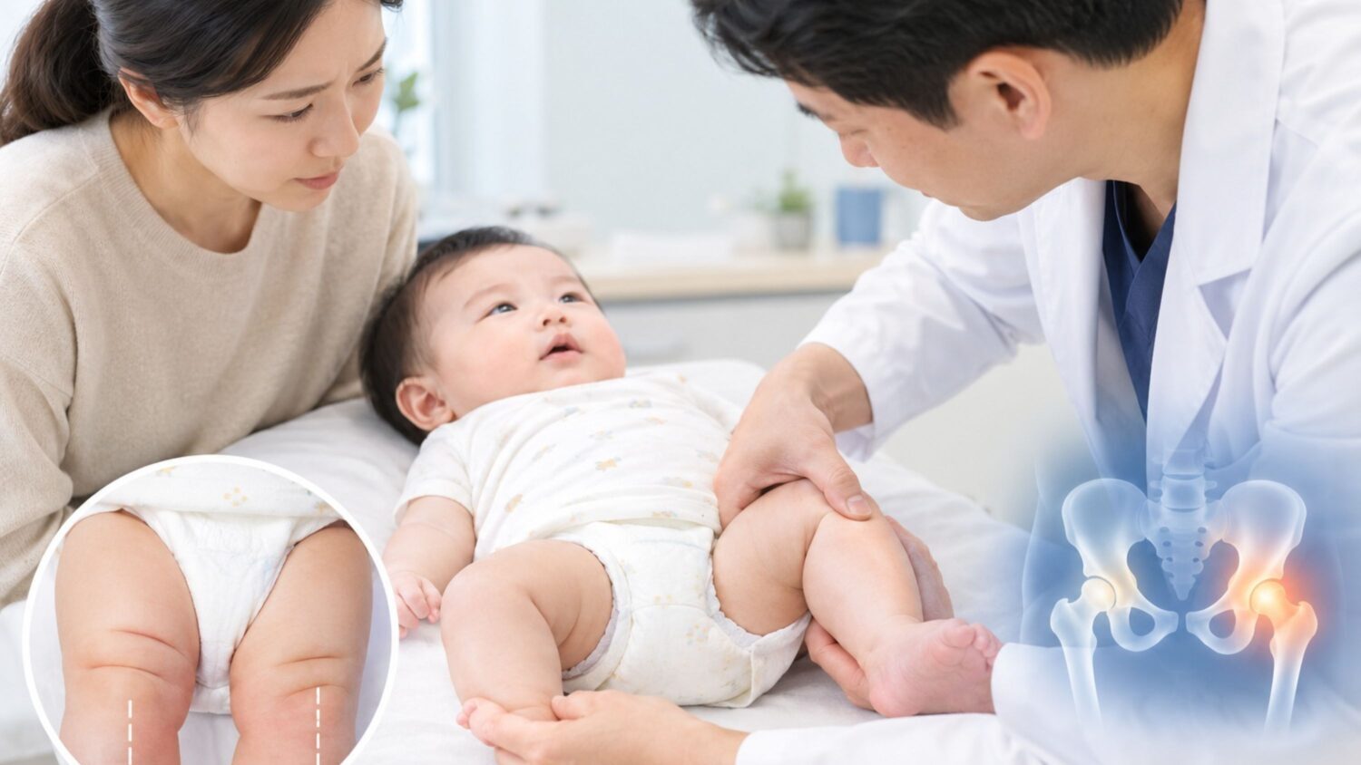
While changing her six-month-old infant's diaper, a parent identified as Kim noticed something subtle yet unusual: the skin creases on her baby’s thighs were asymmetrical, and the right leg did not open as wide as the left when moving the legs apart. The baby showed no signs of discomfort or pain. Upon visiting the hospital, Kim learned her child had developmental dysplasia of the hip (DDH).
Developmental dysplasia of the hip occurs when the femoral head—the rounded top of the thighbone—and the acetabulum, or hip socket, fail to develop properly or fit together securely. The condition may be present at birth or manifest gradually as a child grows. Because it rarely causes pain or striking physical deformities in infancy, careful observation by parents and caregivers plays a pivotal role in early detection.
Key physical indicators include asymmetrical thigh or buttock creases, a noticeable difference in leg length, and restricted mobility in one hip when spreading the legs outward. Once a child begins walking, an uncorrected dislocation may present as a limp or a leaning gait.
However, asymmetrical creases can also occur in healthy infants, meaning they are not a definitive standalone diagnostic tool. Conversely, some babies with DDH show no visible outward signs at all. Primary risk factors include breech birth, a family history of hip dysplasia, female sex, firstborn status, and tight swaddling that forces the legs into a straight, rigid position.
Diagnostic Approach and Age-Specific Treatments
Diagnostic methods vary depending on the infant's age. In newborns and young infants, whose bones remain largely cartilaginous, hip ultrasound is the standard method to visualize cartilage and evaluate joint stability. Once bone formation progresses—typically after 4 to 6 months of age—pelvic X-rays are used to assess bone alignment and socket development.
Treatment strategies depend on the child's age and the severity of joint instability. When caught early, a dynamic harness or brace is typically used to hold the hip in a stable position as the joint develops, though mild cases may simply be monitored closely. If diagnosis is delayed or conservative treatment proves insufficient, specialists may perform a closed reduction under anesthesia followed by spica casting. In severe or late-diagnosed cases, surgical intervention, such as an open reduction or osteotomy, may be necessary.
Even following successful treatment, routine follow-up examinations are critical to monitor joint development and check for potential complications, such as avascular necrosis (impaired blood supply to the femoral head), reducing the risk of early-onset osteoarthritis later in life.
"If a baby's leg creases appear asymmetrical or if one leg seems less flexible when spreading the hips, parents should not dismiss it as a temporary phase of growth," said Lee Dae-hee, head of the Pediatric Orthopedics Center at Ulsan Elijah Hospital. "A prompt medical evaluation is essential, especially if the infant was born in a breech position or has a family history of hip conditions."