

A patient undergoes a magnetic resonance imaging (MRI) scan and is told that their spinal canal has "significantly narrowed" or that a "large herniated disc is pressing on a nerve." Hearing such descriptions, many assume immediate surgery is unavoidable.
While MRI scans play a critical role in spine care, surgeons do not recommend surgical intervention solely because an image looks severe. Other essential clinical criteria determine whether an operation is truly necessary.
"Before deciding on surgery, the primary step is to accurately determine whether a patient's condition genuinely requires surgical treatment," said Park Man-kyu, director of Hu Hospital Busan and head of the International Spine Endoscopy Education Center.
Park is a spine surgeon who trains domestic and international medical professionals in unilateral biportal endoscopy (UBE). The minimally invasive procedure uses two small portals—one for an endoscope and another for surgical instruments—to precisely target and remove lesions while minimizing incision size, tissue damage, and recovery time.
South Korea, particularly the Busan region, has pioneered advanced UBE applications, exporting the technique worldwide. Since 2024, more than 140 international physicians have traveled to Busan to learn under Park’s guidance.
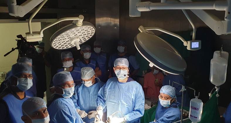
When instructing visiting surgeons, Park highlights one fundamental lesson: "A skilled UBE surgeon is not simply someone who performs a high volume of procedures, but someone who can accurately identify which patients need surgery and which do not."
Evaluating Clinical Symptoms Over MRI Images
Despite rapid advancements in UBE expanding treatable conditions, Park evaluates three clinical indicators before considering an MRI: progressive leg weakness, functional walking capacity, and symptom correlation with imaging findings.
1. Assessing Leg Strength and Neurological Deterioration
In lumbar spinal stenosis—a degenerative condition where a narrowed spinal canal compresses nerves, causing leg numbness and pain—Park prioritizes checking for motor weakness. Rapidly declining leg strength, such as an inability to properly lift the ankle or toes, signals acute neurological impairment.
Severe pain alone does not equate to declining nerve function. If muscle strength remains intact, conservative measures—including medication, therapeutic injections, physical therapy, or exercise—should be prioritized. Conversely, progressive muscle weakness warrants immediate surgical consideration, even if pain is mild.
2. Evaluating Walking Endurance and Daily Function
Spinal stenosis frequently causes neurogenic claudication—pain or numbness triggered by walking that eventually forces a person to stop. When evaluating surgery, Park measures how significantly a patient's walking endurance has declined compared to their baseline. A reduction in walking capacity from 30 minutes down to 5 or 10 minutes, or an inability to complete daily tasks like grocery shopping, commuting, or going out, represents a meaningful functional decline.
If a patient retains good mobility and manages daily routines comfortably despite significant narrowing on an MRI, surgery can be deferred. The same conservative-first approach applies to lumbar disc herniations, which often affect younger adults in their late 20s or 30s. Protruding disc material frequently undergoes spontaneous disc resorption over time. However, surgical intervention becomes necessary if foot drop develops or if unmanageable pain prevents walking, working, or sleeping despite extensive conservative care.
3. Correlating Physical Symptoms with MRI Findings
MRIs frequently reveal degenerative changes across multiple spinal levels, such as simultaneous narrowing at L3–L4, L4–L5, and L5–S1. However, surgeons do not perform multi-level preventive operations simply because multiple segments appear narrowed.
Park emphasizes matching specific symptoms—such as the precise location of leg numbness, sensory loss, or motor deficits—to the corresponding nerve root on the MRI. If symptoms trace back to a single segment, treatment is confined strictly to that targeted area. When diagnostic ambiguity exists, diagnostic tools like selective nerve root blocks, electromyography, or nerve conduction studies are utilized to pinpoint the true pain generator.
Ultimately, effective spine care relies on asking three foundational questions: Is leg strength declining? How severely is walking impaired? Do physical symptoms align with the MRI?
Frequently Asked Questions
Does a severe MRI result mean surgery is unavoidable?
No. MRI findings alone do not determine whether surgery is necessary. Doctors decide based on whether leg strength has declined, how much difficulty the patient has walking or managing daily activities, and whether the actual symptoms correspond to the lesion seen on the MRI.
If pain is severe, is it time for surgery?
Not necessarily. If pain is controlled with medication, injections, exercise, or rehabilitation and nerve function remains intact, continuing conservative treatment is appropriate. Conversely, even if pain is not severe, surgery should be considered more actively when muscle weakness is progressing.
If an MRI shows narrowing at multiple levels, will surgery be more extensive?
Not automatically. Doctors must first identify exactly which segment is causing symptoms. Even when several areas look narrowed, treatment is typically limited to the specific area believed to be causing the patient's discomfort.
What warning sign indicates a medical emergency?
Seek care immediately if leg strength suddenly drops or rapidly worsens, or if bowel or bladder function changes or sensation in the perineal area decreases. Cauda equina syndrome is a neurologic emergency in which multiple peripheral nerve roots are compressed at once. Delayed treatment can result in permanent nerve damage and serious complications, including leg paralysis, sexual dysfunction, and loss of bowel and bladder control.
Park Man-kyu is the director of Hu Hospital Busan and head of the International Spine Endoscopy Education Center. He graduated from Kyungpook National University School of Medicine, completed his neurosurgery residency at Kyungpook National University Hospital, and served as a clinical fellow. Specializing in unilateral biportal endoscopic (UBE) surgery, he has authored 11 chapters across five spine textbooks, including Springer’s 2025 textbook "Unilateral Biportal Endoscopic Spine Surgery: Basic and Advanced Technique," and serves as a peer reviewer for "World Neurosurgery" and the SCI-indexed journal "Neurospine."