
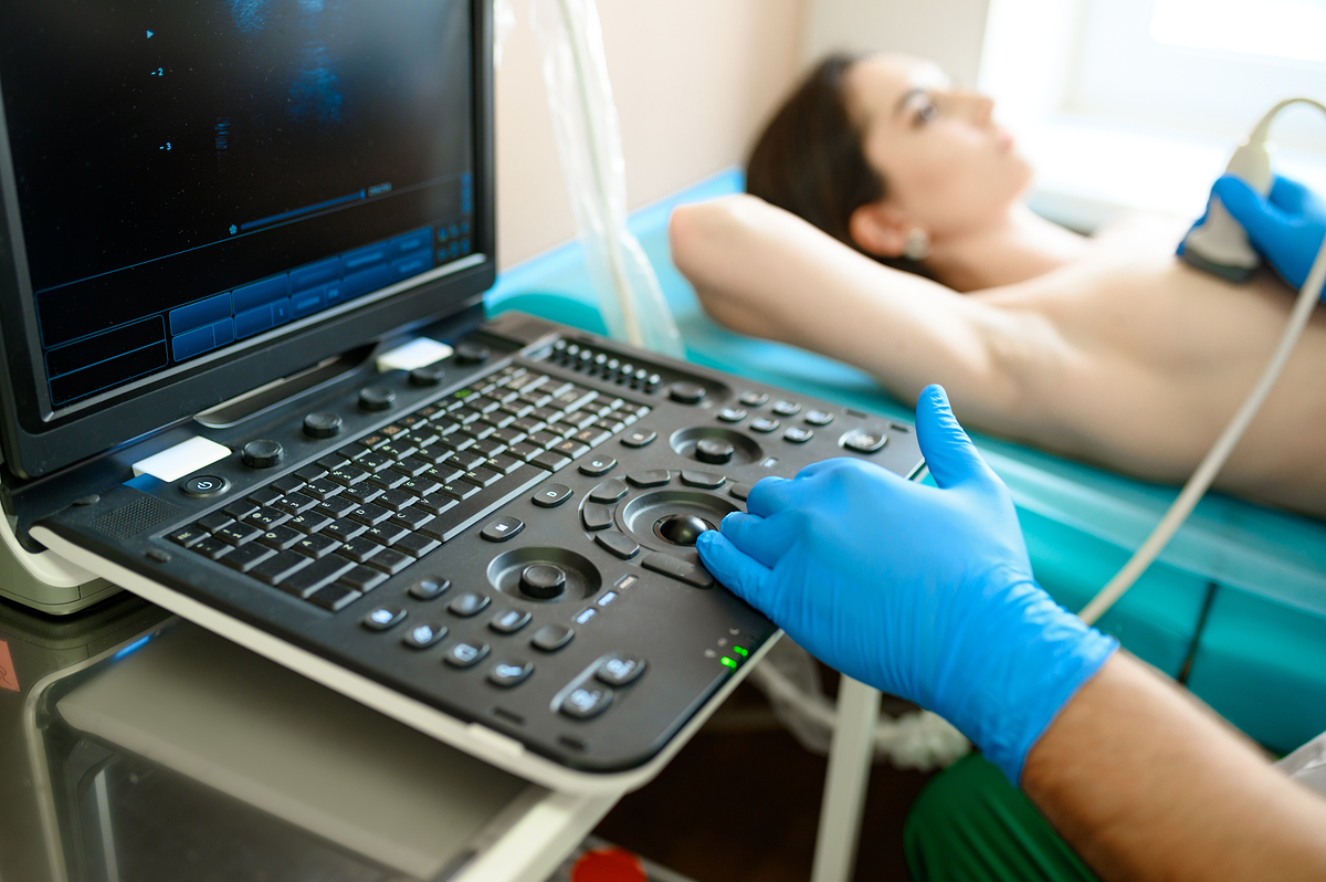
While lying on the ultrasound examination table, hearing the doctor say, "I see a lump here," instantly triggers anxiety. Thoughts race to the worst-case scenario: Is it cancer? Do I need a biopsy? Wouldn't it be safer to just have it removed immediately?
However, not every breast lump requires testing or surgical removal. Updated clinical recommendations jointly issued by the Korean Breast Cancer Society and the Korean Association of Breast and Thyroid Surgeons state that for lesions confirmed as benign via biopsy—where imaging and pathological findings agree—regular follow-up observation is the standard protocol.
Under 2% Malignancy Risk: The "Six-Month Follow-Up" Protocol
Detecting a lump on imaging does not automatically lead to a biopsy. Radiologists and surgeons first evaluate the lesion's shape, margins, orientation, and internal echo patterns using ultrasound to assess the likelihood of malignancy.
"If a lump exhibits classic benign characteristics—such as a typical simple cyst or fibroadenoma—we observe it first," explains Dr. Kim Byung-hyung, Director in the Department of Surgery at Dongnae Bongsaeng Hospital. "Solid masses that are oval, smoothly marginated, and oriented parallel to the skin, as well as certain complex cysts, carry a malignancy risk of less than 2%. In these cases, we perform a follow-up ultrasound six months later."
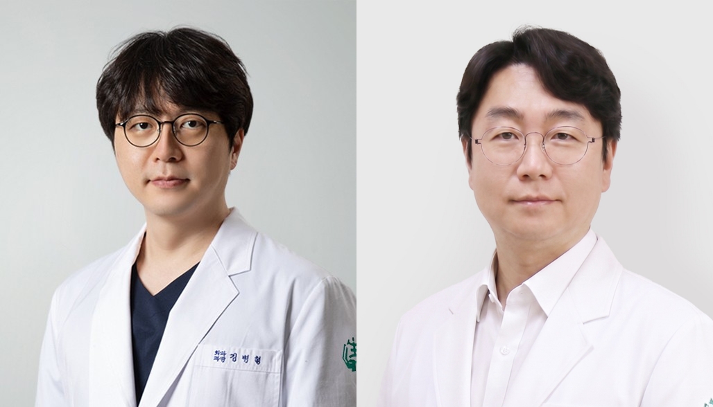
Physicians proceed with a biopsy when suspicious indicators arise. "We perform a biopsy if malignancy is suspected based on irregular shapes, non-parallel orientation, ill-defined margins, architectural distortion, or microcalcifications," Dr. Kim adds. "Immediate tissue sampling is also warranted if a lump exhibits rapid growth, causes bloody nipple discharge, or leads to skin retraction."
Patients undergoing observation should remain alert to physical changes. Key warning signs include noticeable growth or hardening of the lump, changes in skin or nipple appearance, abnormal discharge, or a new palpable mass in the armpit.
When Benign Lumps Warrant Removal
If a biopsy confirms a benign lesion that aligns with ultrasound findings, routine monitoring remains the standard approach. However, surgical or vacuum-assisted removal may be considered under specific circumstances:
Persistent pain or local discomfort
Physical discomfort or psychological distress caused by a palpable mass
Cosmetic concerns regarding breast contour
Significant, documented increase in lesion size
If a patient expresses a strong desire for removal after a comprehensive clinical consultation, excision remains a valid option. In such cases, the key clinical factor is not merely the presence of a lump, but its precise pathological profile.
Mammotome: Getting the Diagnostic Sequence Right
Vacuum-assisted excision (VAE), widely known by the brand name "Mammotome," is a minimally invasive procedure that uses a specialized thick needle and vacuum suction to remove breast tissue under ultrasound guidance. Updated guidelines emphasize that the critical issue is not the procedure itself, but the sequence of care. Experts caution against the preemptive approach of removing a mass before establishing a definitive diagnosis.
Standard protocol dictates identifying the lesion first through core needle biopsy (CNB) or vacuum-assisted biopsy (VAB) before deciding on removal. Proceeding directly to VAE without a prior biopsy is restricted to select cases where imaging demonstrates unambiguous benign features.
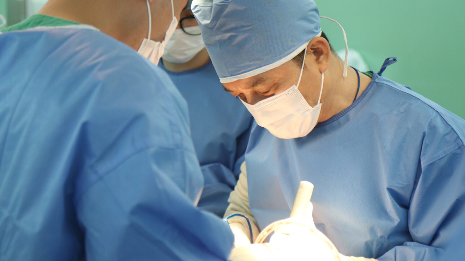
VAE serves specific clinical utility when applied appropriately. "VAE is particularly suitable when a patient wishes to completely remove a small lesion previously confirmed benign on biopsy, avoiding open surgery," says Dr. Lee Sang-min, Director in the Department of Surgery at Dongnae Bongsaeng Hospital.
Dr. Lee notes that VAE is frequently considered for benign masses measuring between 1.5 cm and 2 cm before further enlargement occurs. While the procedure can be performed on smaller or larger lesions, decisions are made by weighing clinical judgment alongside patient preference.
However, the medical approach changes entirely if malignancy is suspected. Guidelines state that standard surgical excision must be prioritized to establish a accurate diagnosis and formulate an oncologic treatment plan. VAE is not recommended as a curative treatment for suspected or confirmed breast cancer, nor can it substitute for standard oncologic surgery.
Same-Day Diagnostics, Scheduled Excision
For patients who discover a lump, navigating the clinical process efficiently is paramount. At specialized centers like Dongnae Bongsaeng Hospital, patients can undergo physical examination, mammography, and ultrasound on the same day as their initial consultation, with the attending surgeon reviewing ultrasound images directly to explain the findings.
When tissue sampling is required, core needle biopsies are routinely performed during the initial visit. Simple procedures, such as aspirating fluid from symptomatic cysts, are also completed immediately during the ultrasound exam.
Scheduling for VAE depends on the diagnostic path. "If ultrasound reveals a clearly benign lesion, VAE can occasionally be performed on the same day under local anesthesia," Dr. Lee explains. "However, if a biopsy is required, pathology results take approximately one week. In those instances, any subsequent excision is scheduled for a separate date."
Frequently Asked Questions After a Breast Exam
Q. If I have dense breasts, do I always need a breast ultrasound?
A. Dense breast tissue is a common anatomical variation, not a disease. However, because dense tissue can obscure lesions on standard mammography, supplemental ultrasound is often recommended based on individual risk factors and mammographic findings.
Q. If I feel a lump in my breast, should I get a mammogram or an ultrasound first?
A. The choice depends on patient age, clinical symptoms, and lesion location. Because mammography and ultrasound evaluate different tissue characteristics and complement each other, both exams are frequently performed together.
Q. Does finding a lump on an ultrasound mean a biopsy is mandatory?
A. No. Lesions with a very low likelihood of malignancy are monitored at fixed intervals. Biopsies are reserved for lesions displaying suspicious features or significant changes over time.
Q. Does a growing benign lump always have to be removed?
A. Size alone does not dictate treatment. Surgeons evaluate the rate of growth, presence of symptoms, cosmetic considerations, and any changing features on imaging before recommending removal.
Q. Is a Mammotome procedure the same thing as a biopsy?
A. No. Core needle biopsy (CNB) and vacuum-assisted biopsy (VAB) are diagnostic tests designed to extract small tissue samples for pathology. Vacuum-assisted excision (VAE or Mammotome) is a therapeutic procedure intended to remove a lesion entirely. Although VAB and VAE may utilize similar vacuum-assisted equipment, their clinical objectives differ.