


Soccer player Park Ji-sung and marathoner Lee Bong-ju both have flat feet. Lee even had mismatched feet, with his left foot roughly 4 millimeters larger than his right, yet he ran longer and farther than almost anyone.
So are flat feet really a problem? Do they inevitably make walking or running for long periods difficult, and do they cause trouble as you age?
Not necessarily. Some people have had flat feet on both sides since childhood, while others have normal feet until adulthood, when the arch in one foot begins to collapse. While they may look similar on the surface, they are fundamentally different conditions.
The latter should never be dismissed as "just flat feet." As the arch collapses and the alignment of the heel and forefoot shifts, pain can worsen, making even routine walking difficult in severe cases.
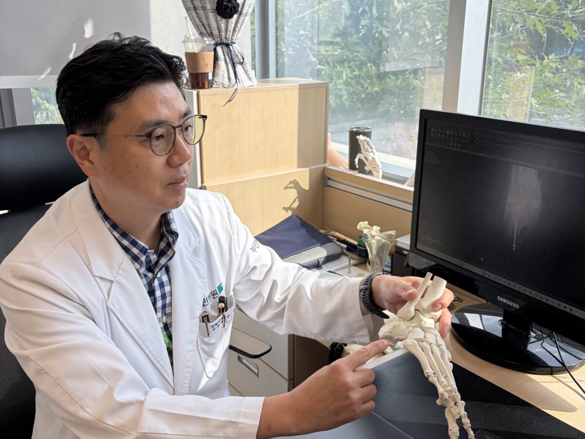
Speaking on August 30 at the Korean Foot and Ankle Society symposium held at the Samsung Medical Center Cancer Hospital auditorium, Ahn Ji-yong, director of the Foot and Ankle Joint Center at Seoul Bumin Hospital, addressed "Adults with persistent pediatric flatfoot vs. adult flatfoot: How is treatment different?" His central message was clear: "Accurate differentiation between the two types of flatfoot is crucial."
"Adult-acquired flatfoot," which develops in adulthood, has recently been reframed in medical literature as Progressive Collapsing Foot Deformity (PCFD). It does not simply mean having a low arch; it refers to a complex structural deformity in which the alignment of tendons, ligaments, and bones collapses as a whole. Crucially, without proper treatment, the condition continuously progresses over time.

If Both Feet Have Been Flat Since Childhood, It Is Not Necessarily a Disease
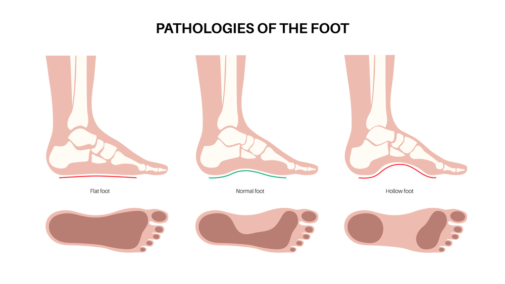
Flatfoot is a condition where the medial longitudinal arch along the inside of the sole is lower than normal or nearly absent during weight-bearing. It is far more common than many realize. In a study of Korean adults, 22.1% met the radiologic criteria for flatfoot based on talo-first metatarsal angle measurements, even though only 8.8% of participants reported believing they had flat feet. In short, many people live without realizing they have flat feet.
Among them, "flexible flatfoot" that persists from childhood typically affects both feet symmetrically. Even with a low arch, the surrounding bones, ligaments, and tendons adapt over time to maintain structural balance. Ahn described this as "a stable structure that maintains balance in a lowered state."
In such cases, if there is no significant pain or functional impairment, treatment is not required. This structural adaptation is why athletes like Park and Lee were able to perform at an elite level despite having flat feet.
This does not mean issues will never arise. A low arch alters load-bearing forces and places different stresses on tendons and ligaments, making the feet and ankles tire more easily during prolonged standing or walking. Other deformities may also develop after middle age due to shifting weight distribution, though lifelong flatfoot itself is not the direct cause.
If the Arch in One Foot Drops After Your 40s or 50s, That Is Where the Story Changes
Flatfoot becomes a serious medical concern when a previously normal arch in one foot begins to lower gradually after middle age, signaling a progressive deformity across the entire foot.
In many cases, the condition begins with degeneration of the posterior tibial tendon—the key tendon supporting the inner arch. As the supporting ligaments weaken, the heel tilts outward, the forefoot spreads, and overall foot alignment progressively deteriorates.
Early on, patients may experience aching along the inside of the ankle and fatigue after long walks. However, as the deformity advances, the pain shifts to the outside of the foot due to bone impingement. Patients often mistake the disappearance of inner ankle pain for improvement, but it actually indicates that the condition has progressed to a more severe stage.
This is why Ahn urged primary-care orthopedists to confirm two fundamental questions: "Since when has your arch become this low?" and "Is it affecting both feet, or only one?"
Ahn noted that these two questions constitute "half the diagnosis." If both feet have been flat since childhood, it is likely persistent flexible flatfoot. But if only one foot has flattened noticeably over the past one to two years, clinicians must suspect adult-acquired flatfoot. Even if the feet look similar externally, their underlying pathology and treatment approaches are completely different.
Left Untreated, Even Walking Becomes Difficult: Why It Is a 'Race Against Time'
As PCFD continues to progress, it impairs the ability to walk. The collapse in alignment reduces the foot's efficiency in supporting body weight and pushing off the ground.
Discomfort that once occurred only after long walks turns into fatigue even on flat ground. Rising onto tiptoes becomes difficult, pain worsens as outer foot structures impinge on one another, and the foot gradually stiffens, severely impacting daily life.
For this reason, treatment is a race against time. While the foot remains flexible, joint-preserving corrective options are available. Once the deformity progresses to a rigid stage, treatment choices shrink dramatically.
In early stages, management focuses on reducing pain and slowing progression. If posterior tibial tendon inflammation is severe, patients may use a walking boot or short-leg cast for temporary rest, followed by bracing, strengthening exercises, and balance training. However, if pain persists or the condition worsens after 3 to 6 months of conservative management, surgery should be considered. Delayed diagnosis complicates treatment, often requiring complex reconstructive surgery involving tendons, ligaments, and bones.
If the deformity reaches a rigid stage that cannot be corrected manually and secondary arthritis develops in joints like the subtalar joint, procedures such as subtalar arthrodesis or double/triple arthrodesis may be necessary. If the deformity extends to the ankle joint and the joint surface tilts outward, deltoid ligament reconstruction may be considered; if joint stiffness or advanced arthritis is present, clinicians may evaluate ankle arthrodesis or total ankle replacement.
Don't Start with Shoe Insoles—Get a Weight-Bearing X-Ray First
If your foot shape changes in adulthood, confirming the underlying cause must take priority over purchasing functional shoe insoles. In many instances, the arch appears normal while sitting or lying down and collapses only under weight-bearing conditions.
The essential first step is obtaining weight-bearing foot and ankle X-rays to assess arch height, heel alignment, and bilateral differences. If necessary, MRI or weight-bearing CT scans should be used to evaluate posterior tibial tendon damage, ligament integrity, and joint alignment.
[Checklist] “So what about my feet?” … the differences between the two types of flatfoot at a glance
| Take a look | Yes | No |
| Even in childhood photos, were both feet flat like they are now? | □ | □ |
| Over the past few years, has only one foot become flatter? | □ | □ |
| Has the sole of only one shoe started wearing down noticeably differently? | □ | □ |
| Does it hurt or swell when you press behind the inside of the ankle bone? | □ | □ |
| From behind, does one heel tilt outward noticeably more than the other? | □ | □ |
| Is it hard to rise onto tiptoe on one foot, or does it hurt if you repeat it several times? | □ | □ |
| Your inner ankle used to hurt — has pain recently started on the outside as well? | □ | □ |
Experiencing a few of these symptoms does not automatically confirm PCFD. However, if one foot is gradually collapsing in adulthood alongside pain and walking difficulty, it should never be dismissed as "just flat feet." The critical distinction lies not in how low the arch is, but whether it has remained stably low since childhood or is newly collapsing in adulthood. While they may appear similar, a stably low arch and a progressive foot deformity are fundamentally different medical conditions.