
When pain or numbness in the buttocks and legs persists after spinal surgery, it does not automatically indicate that the operation was a failure. Instead, the culprit may be a hidden lesion in the narrow passageway where nerves exit the spinal column.
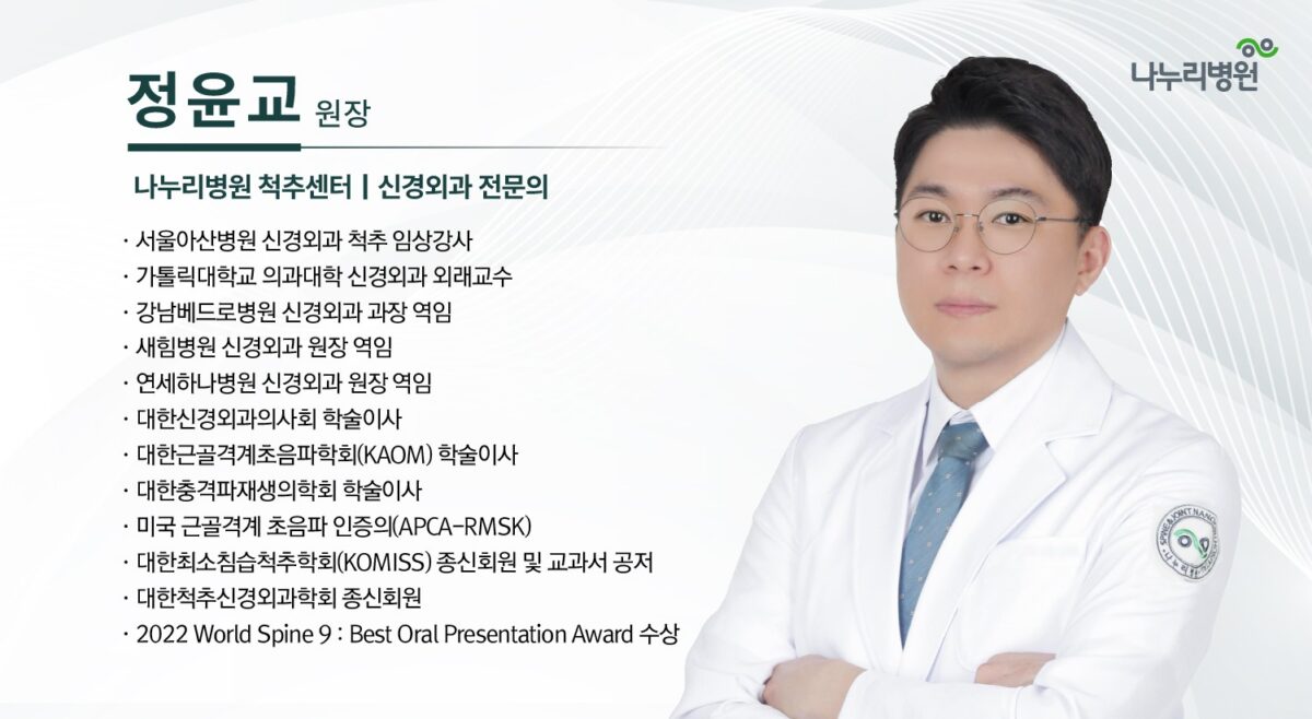
Jeong Yun-gyo, director at the Spine Center at Nanuri Hospital, presented diagnostic strategies and endoscopic treatment approaches for foraminal lesions on the 23rd at the 2026 Korean Neurosurgical Hospital Association's 12th Academic Conference, held at Sejong University's Gwanggaeto Hall in Seoul. Delivering a lecture titled "Foraminal Pathology Unlocked: Case for Endoscopic Access," Jeong detailed methods for identifying these obscure lesions and reaching them via endoscopic techniques.
Understanding the Nerve 'Exit' and the Hidden Zone
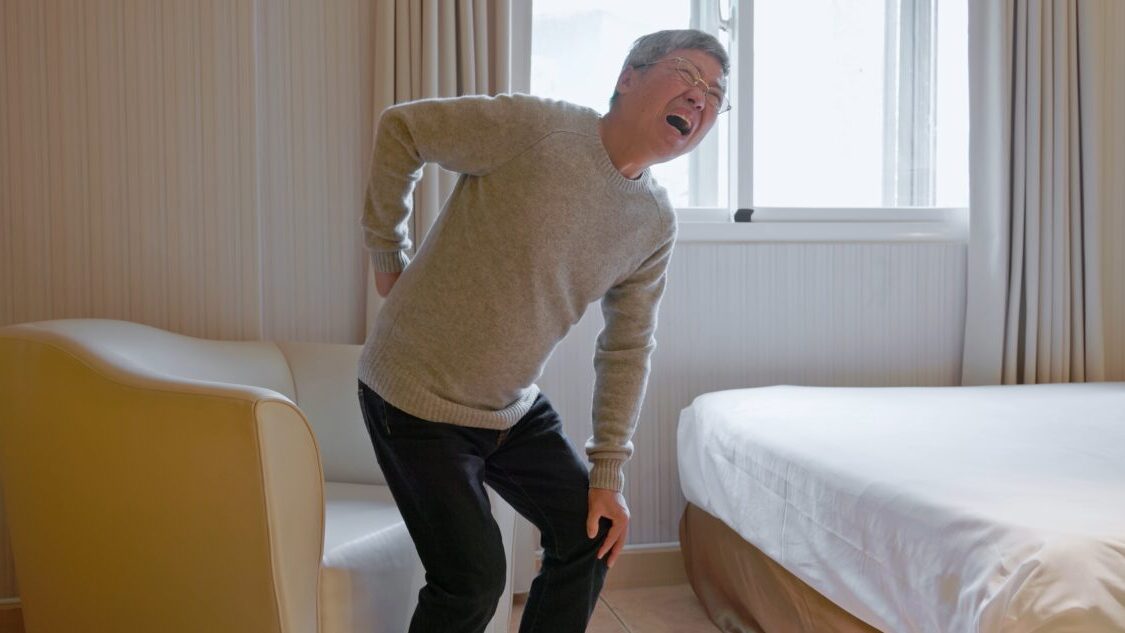
Spinal nerves branch off from the main spinal cord and exit the column through small lateral openings between adjacent vertebrae known as the intervertebral foramina. With age, reduced disc height and the thickening of surrounding joints and ligaments can narrow these openings, causing foraminal stenosis. When a nerve becomes compressed in this exit, pain or numbness can radiate from the lower back down through the buttocks, thighs, and calves.
Identifying these lesions poses a challenge because the structures surrounding the intervertebral foramen sit deep within the spinal architecture. The area where the inner portion of the foramen meets the side of the spinal canal is particularly difficult to visualize or reach with standard surgical instruments—a region spine surgeons often refer to as the "hidden zone."
Rethinking Post-Surgical Pain Beyond 'Failed Surgery'
Persistent or recurring pain following spinal operations was historically categorized under the broad label of "Failed Back Surgery Syndrome." However, the term can be misleading, as it implies the initial surgery went wrong when the actual underlying causes vary widely.
Post-surgical pain can stem from recurrent disc herniation, epidural scar formation, spinal instability, or newly compressed nerves in adjacent areas. Inadequately addressed foraminal stenosis from the primary surgery is another key factor requiring evaluation. Rather than rushing into revision surgery, clinicians must reassess the patient's symptoms, physical examinations, and pain triggers from the ground up.
Diagnosing foraminal lesions also requires looking beyond MRI scans. Advanced imaging can show structural narrowing that does not correspond to a patient's pain, or conversely, fail to capture clear abnormalities despite severe symptoms. As a result, clinicians must evaluate sensory loss, muscle weakness, and postural changes alongside imaging to confirm the exact source of nerve compression.
Targeted Treatment Through Spinal Endoscopy
To address these deep-seated lesions, Jeong presented real-world cases demonstrating the efficacy of spinal endoscopy. Using thin endoscopes and specialized instruments inserted through minimal incisions, surgeons can directly visualize and decompress the nerve while leaving surrounding muscle and joint structures largely intact.
Jeong also addressed complex anatomical challenges, such as accessing the L5-S1 segment—where the lowest lumbar vertebra meets the pelvis—which is frequently obstructed by the iliac crest and surrounding pelvic structures.
Despite its benefits, spinal endoscopy is not a universal solution. Patients with extensive structural stenosis or spinal instability may require alternative surgical approaches.
"The intervertebral foramen is a critical neural pathway, but its depth makes lesions easy to overlook," said Jeong Yun-gyo. "In endoscopic spinal procedures, accurate diagnosis and a precise, targeted approach are essential to resolve the compression while minimizing damage to healthy tissue."
Jeong added, "For patients with persistent post-surgical symptoms, re-evaluating the pain source with precision is crucial. We will continue to build on clinical data and technical expertise to deliver more refined, minimally invasive care."