

Researchers have identified key genetic markers that could help estimate the risk of recurrence in esophageal cancer patients who have residual tumor tissue following treatment. In surgical samples collected after preoperative chemoradiotherapy, heightened activity in inflammation-related gene sets was associated with significantly higher risks of disease progression, recurrence, and death.
A Seoul National University Hospital research team—led by Radiation Oncology Professors Jang Beom-seop and Kim Hak-jae, Cardiothoracic Surgery Professor Na Kwon-jung, and Pathology Professor Lee Hye-seung—reported these findings after analyzing surgical tissue from 56 patients with esophageal squamous cell carcinoma. Using "spatial transcriptomics"—an advanced molecular profiling technique that measures gene activity while preserving precise location information within tissue—the team created a molecular map of post-treatment tumors. The study was published in the international scientific journal npj Precision Oncology.
Inflammatory Gene Activity Linked to a 2.5-Fold Increase in Risk
In clinical practice, predicting long-term outcomes for patients classified with pathologic regression grades 1 to 2—indicating that substantial cancer tissue remains after chemoradiotherapy—remains challenging based on tissue appearance alone.
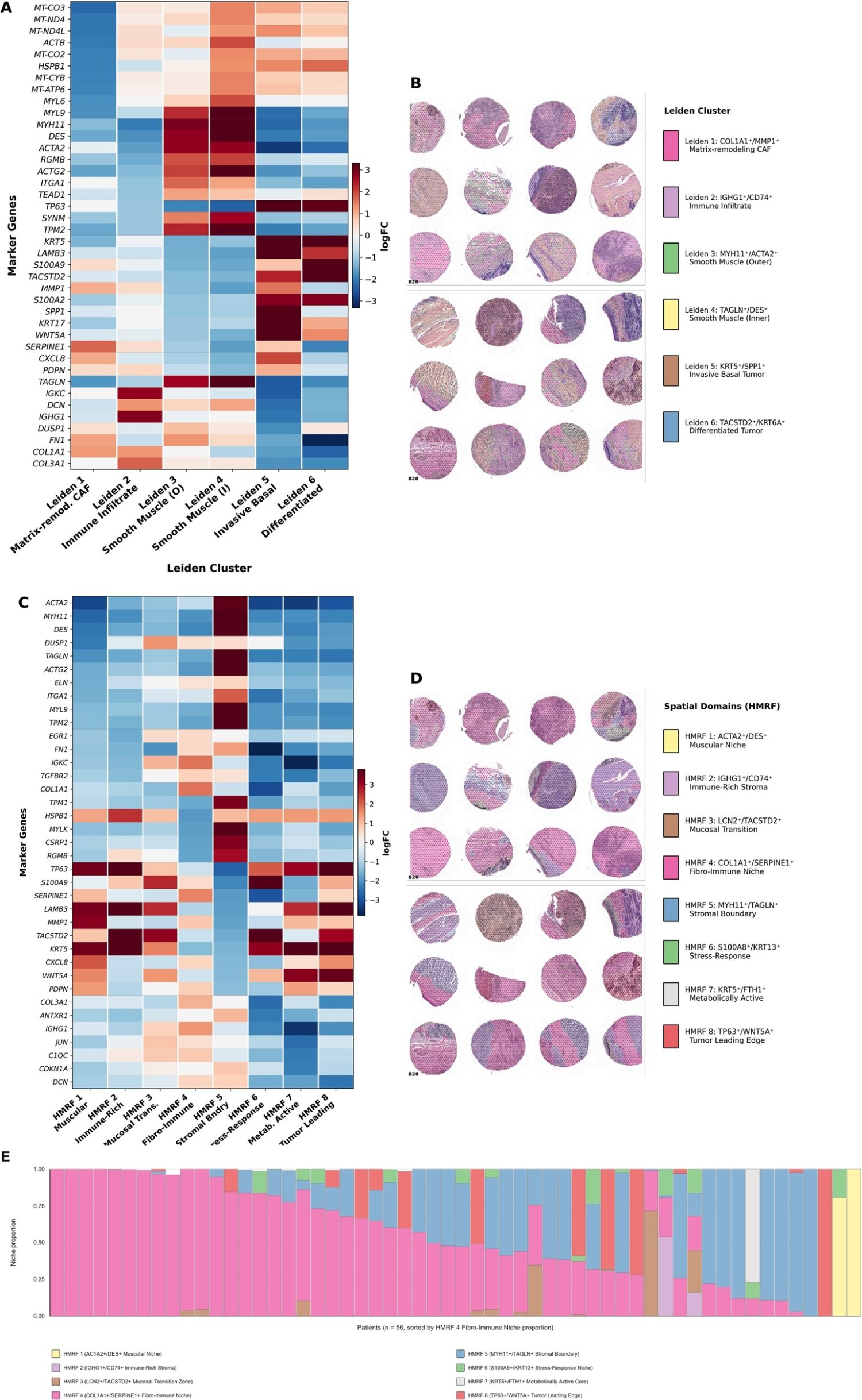
To address this limitation, the research team performed a targeted analysis on 27 patients whose tumor tissue was predominantly composed of a "fibro-immune niche" (accounting for more than 50% of the cancer tissue). Within this subgroup, every one-standard-deviation increase in the inflammatory gene-set score was associated with a 2.53-fold increase in the combined risk of progression, recurrence, and death.
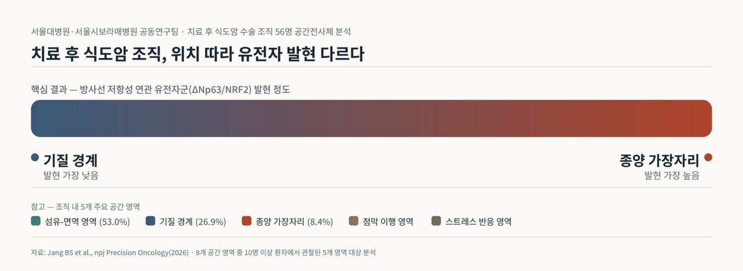
By categorizing the tissue microenvironment into eight distinct niches, the researchers also mapped the spatial distribution of the "ΔNp63/NRF2" gene set, which is cited as potentially linked to radioresistance. This gene set reached its highest expression levels at the tumor edge and its lowest at the stromal boundary, highlighting how therapeutic resistance can vary significantly across different regions within the same tumor tissue.
Early Findings Lay Groundwork for Precision Oncology
Because this was an initial exploratory study analyzing post-treatment surgical tissue, the researchers noted that the findings cannot yet be applied directly to routine bedside care. Furthermore, the risk metrics calculated from the 27-patient subgroup represent group-level statistical trends rather than individual prognostic scores. The identified gene sets serve as candidates for future clinical validation.
"This study provides a meaningful foundation for understanding the complex post-treatment microenvironment of esophageal cancer," said Professor Jang Beom-seop. "It will serve as a vital stepping stone for advancing future precision-medicine research."