

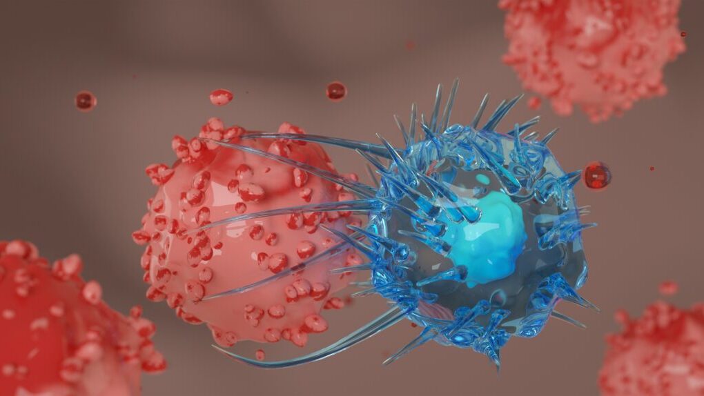
Some immune cells surrounding a tumor actively attack it, while others create conditions that help it survive. A South Korean research team has discovered that the strength of the body’s anti-cancer response varies sharply depending on which immune cells reach the tumor site first.
The study focused on glioblastoma, a malignant brain tumor known for its aggressive nature, high recurrence rate, and resistance to treatment. When natural killer (NK) cells—which directly attack cancer cells—arrived at the tumor first, the anti-cancer response was significantly enhanced. Conversely, when microglia—immune cells native to the brain—arrived first, the attack capability of NK cells was markedly weakened.
A joint research team led by Prof. Baek Sun-ha and Dr. Kim Yo-na of the Department of Neurosurgery at Seoul National University Hospital, alongside Prof. Park Sung-soo and Dr. Han Seok-gyu of the Department of Mechanical Engineering at Sungkyunkwan University, announced the findings on July 30 after developing an experimental microfluidic device that controls the sequence of immune cell entry into a tumor.
Same cells, different outcomes when arrival order changes
Glioblastoma develops in the brain, growing rapidly and infiltrating surrounding brain tissue. Even with a combination of surgery, radiation therapy, and chemotherapy, frequent recurrence makes complete treatment exceptionally difficult.
One major factor complicating treatment is the immune microenvironment surrounding the cancer. While microglia typically clear damaged tissue and foreign material in the brain, they can adapt near glioblastoma cells to create conditions favorable for cancer survival. NK cells, by contrast, actively seek out and destroy cancer cells or virus-infected cells.
The researchers investigated whether the sequence in which these two immune cell types encounter cancer cells could fundamentally alter the tumor’s response.
Because conventional cell-culture methods make it difficult to control when specific cells arrive at a tumor site, the research team constructed a microfluidic device capable of introducing selected cells through multiple channels in a precise, predetermined order.
Using this device, they created spherical 3D artificial brain tumor tissues roughly 400 micrometers in diameter. Holding the overall proportions of cancer cells, microglia, and NK cells constant, the only variable was the arrival sequence of the cells.
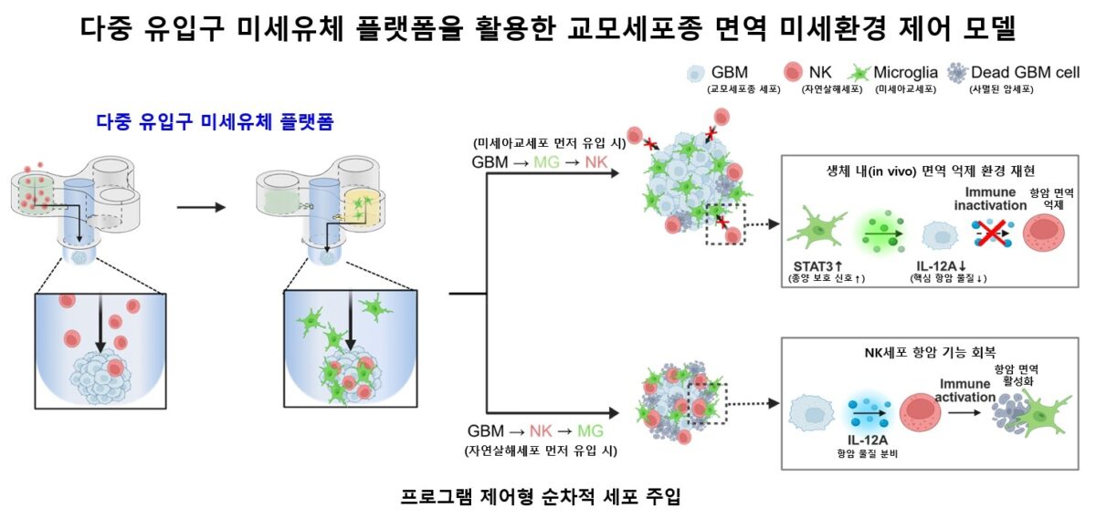
The results were clear. When microglia entered first, "STAT3" signaling—which protects cancer cells and suppresses immune activity—became significantly more active. NK cells arriving afterward struggled to penetrate the tumor, and their cancer-killing capability declined.
Reversing the order produced the opposite effect. When NK cells arrived first, genes linked to anti-cancer attacks became highly active. Despite introducing identical cell types and quantities, changing only the arrival sequence fundamentally altered the tumor's immune microenvironment.
Differences observed in tumor cells from patients with varying survival times
Similar structural differences appeared in tumor cells obtained from actual patients.
The researchers compared tumor cells from a patient who survived for more than 10 years with those from a patient with a relatively short survival period. Tumor cells from the short-survival patient proliferated more rapidly when paired with microglia and displayed stronger resistance against NK-cell attacks.
The team also identified the specific genes involved in these differences. A key gene that stood out was "IL12A," which produces a subunit of interleukin-12 (IL-12), a protein that supports anti-cancer immune activity. When NK cells entered the tumor first, expression of IL12A increased markedly.
After analyzing approximately 338,000 cells from 27 glioblastoma patients, researchers detected IL12A in only two patients, accounting for just 0.45% of total cells analyzed. This indicates that this vital immune-activating signal is heavily suppressed in glioblastoma tissue.
Large-scale patient data confirmed the pattern: patients with higher IL12A expression demonstrated significantly longer overall survival times.
Based on these findings, the researchers highlighted IL12A as a potential biomarker for evaluating glioblastoma immune status and predicting patient prognosis.
Can drugs dismantle the cancer’s immune shield?
This raises a key question: Even after microglia establish an early foothold, can the immunosuppressive environment protecting the cancer be reversed?
To test this, the researchers administered WP1066—a drug that blocks the STAT3 signaling involved in immune suppression—together with temozolomide, the standard chemotherapy drug for glioblastoma.
Following treatment, IL12A expression and secretion, which had been suppressed by microglia, increased once again. NK cells were able to penetrate deeper into the tumor tissue, resulting in a stronger cancer-killing effect.
These findings suggest that treatment outcomes could be improved not only by directly targeting cancer cells, but also by reshaping the immunosuppressive microenvironment around the tumor.
However, these therapeutic effects have not yet been confirmed in human clinical trials. This study utilized 3D artificial brain tumor models, patient-derived tumor cells, and existing patient data. Whether combining WP1066 and temozolomide improves real-world clinical outcomes or extends survival must be verified through future clinical trials.
Additionally, the microfluidic device developed by the team is not yet ready for direct diagnostic testing in clinical settings. Nevertheless, it offers a powerful research platform to replicate an individual patient's tumor immune microenvironment in vitro and evaluate treatment responses.

"This study demonstrates that the immune environment of glioblastoma is dynamic rather than fixed, changing based on the sequence in which cancer cells and immune cells interact," Baek said. "This platform will serve as an important breakthrough for predicting treatment responses and establishing personalized immunotherapy strategies for patients with hard-to-treat brain tumors."
The study was supported by the Ministry of Science and ICT and the National Research Foundation of Korea, and the findings were published in the international journal Neuro-Oncology.