

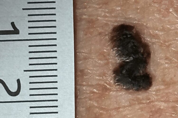
It looked like melanoma, a potentially fatal form of skin cancer, but detailed testing revealed the lesion to be pigmented seborrheic keratosis—commonly known as age spots. This finding was recently highlighted in a case report published in the medical journal Cureus.
Clinicians at the University of Colorado School of Medicine detailed the case, in which a lesion highly suspicious for melanoma was fortunately confirmed to be a benign seborrheic keratosis. According to the medical team, a man in his 60s sought treatment for a dark-brown skin lesion on his upper back. The patient reported that the growth had enlarged slowly over several years and was painless. Upon examination, doctors found a slightly raised, dark-brown patch measuring 1.2 cm x 0.7 cm with an irregular oval shape.
Because of the high clinical suspicion of melanoma, the team surgically removed the lesion and performed a biopsy. The pathology results ultimately confirmed the growth was a seborrheic keratosis rather than a malignancy.
Melanoma is a cancer that originates in melanocytes and is most common on the skin. While it may initially resemble a common mole or dark spot, it is a highly invasive cancer capable of metastasis. Warning signs of melanoma include lesions that grow rapidly, feature irregular borders, contain multiple colors, exceed 6 mm in diameter, or bleed.
In contrast, seborrheic keratosis is a benign growth composed of epidermal keratinocytes. While both moles and seborrheic keratoses can appear brown or black, they develop through different biological mechanisms. A typical mole is formed by a cluster of pigment-producing melanocytes, whereas seborrheic keratosis is a noncancerous growth that becomes more prevalent with age. Their textures also differ: moles are generally smooth patches or small bumps, while seborrheic keratosis tends to have a rough, "stuck-on" appearance.
Distinguishing Benign from Malignant
The clinical team noted that while most large patches are seborrheic keratosis, an excisional biopsy remains necessary whenever melanoma is suspected.
“Another option is dermoscopy to help distinguish benign from malignant lesions,” the clinicians explained. Dermoscopy is a specialized examination that allows doctors to evaluate skin lesions under high magnification and controlled lighting to identify subtle markers of cancer that are invisible to the naked eye.