

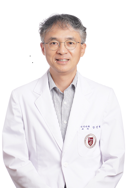
Korea University Anam Hospital announced on Oct. 22 that Professor Kang Shin-Hyuk of its Department of Neurosurgery received an academic award at the 66th Annual Fall Meeting of the Korean Neurosurgical Society, held Oct. 15–17, 2026.
Kang was recognized for leading research on a novel technique combining confocal laser endomicroscopy with artificial intelligence (AI) for rapid intraoperative brain tumor diagnosis. The method applies a fluorescent dye to surgical tissue specimens, captures high-resolution images, and interprets them to generate an immediate diagnosis.
The award-winning paper is titled “AI Augmented Confocal Laser Endomicroscopy for Rapid Intraoperative Diagnosis of Brain Tumors.”
In brain tumor surgery, surgeons must remove tumor tissue as completely as possible while sparing healthy brain tissue. However, frozen-section analysis—the conventional intraoperative diagnostic standard—typically requires 20 to 30 minutes, limiting real-time decision-making in the operating room.
The research was conducted as a prospective multicenter clinical study involving Korea University Anam Hospital, Seoul National University Hospital, Samsung Medical Center, and St. Michael’s Hospital at the University of Toronto in Canada. Kang served as a corresponding author.
The team evaluated the diagnostic accuracy of confocal laser endomicroscopy while developing and validating an image-based AI diagnostic model using 461 tissue samples from 376 patients.
Confocal laser endomicroscopy achieved 94% diagnostic accuracy, matching the performance of frozen-section analysis (92%). The median time to obtain a diagnosis was 5 minutes and 56 seconds—approximately 14 minutes faster than the 20 minutes needed for frozen-section analysis. The AI model demonstrated 94% accuracy in detecting tumor presence and 88% accuracy in classifying tumor types.
“This award is especially meaningful as it recognizes our team’s efforts to achieve faster, more accurate diagnoses during brain tumor surgery by combining artificial intelligence with confocal laser endomicroscopy,” Kang said. “We will continue advancing intraoperative diagnostic technologies to aid clinicians in making precise decisions and improve patient safety and outcomes.”
The study utilized the cCeLL platform, a confocal laser endomicroscopy-based digital tissue biopsy system developed by VPIX Medical. As part of a joint Korea-U.S. research-centered hospital innovation project, the team is currently conducting follow-up studies in collaboration with Korea University Anam Hospital, Korea University’s AI research team, Seoul National University Hospital, Samsung Medical Center, Boramae Medical Center, the National Cancer Center, VPIX Medical, Harvard University, and the Hospital of the University of Pennsylvania.
The findings were published in April in npj Digital Medicine, a journal published by Nature Portfolio.