

Sharp, stinging pelvic pain and dozens of exhausting trips to the bathroom in a single day—these are the debilitating hallmarks of interstitial cystitis, also known as bladder pain syndrome. As the bladder fills with urine, the pain intensifies, easing only temporarily after urination. Yet, standard clinical tests frequently reveal nothing unusual, leaving thousands of patients exhausted as they bounce from hospital to hospital in search of answers.
In a disease that has long lacked clear explanations due to its unknown etiology, a South Korean research team has successfully mapped the hidden network of signals exchanged between cells inside the bladder wall, offering new hope for targeted treatments.
Samsung Medical Center announced on July 16 that a research team led by Professors Kang Min-yong and Ko Kwang-jin of the Department of Urology has identified a complex cell signaling network closely linked to the onset of interstitial cystitis. The groundbreaking study was published on July 3 in the prestigious international journal Experimental & Molecular Medicine.
Debilitating Symptoms and Diagnostic Hurdles
Interstitial cystitis develops when the inner lining of the bladder becomes chronically damaged and inflamed, triggering severe pelvic pain, extreme frequency, and an uncontrollable urgency to urinate. Long classified as one of the most intractable urological conditions, it has historically lacked a fundamental cure because its underlying cause remained unknown. Compounding the challenge, symptoms vary drastically from patient to patient, and traditional endoscopic diagnostics have focused only on surface-level, visible lesions, failing to explain how individual cells drive the progression of the disease.
To uncover these deep cellular mechanisms, the research team analyzed full-thickness bladder tissue samples from eight severe "Hunner-type" patients—individuals with advanced disease characterized by ulcer-like Hunner lesions who ultimately required surgery to remove or enlarge the bladder. For a control group, the researchers obtained healthy bladder tissue from two individuals undergoing pelvic surgery for unrelated reasons. In five of the eight surgical patients, the team was able to perform paired comparisons by extracting both active lesion tissue and visually normal tissue from the same bladder, effectively minimizing patient-to-patient genetic variables.
A Multicellular Network Amplifying Pain
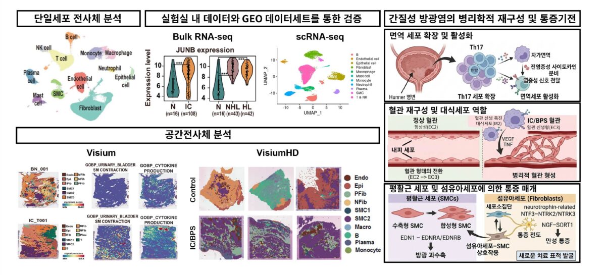
Departing from traditional methods that examine only tiny mucosal biopsies, the team removed intact, full-thickness sections of the bladder wall down to the deeper muscular layers. They combined this comprehensive tissue sampling with two cutting-edge technologies: single-cell transcriptomics, which reads the genetic expression of individual cells, and high-resolution spatial transcriptomics, which pinpoints exactly where these cells reside within the tissue architecture.
The analysis revealed a complex "multicellular network" where stromal connective tissue cells, immune cells, and vascular cells are intricately intertwined and constantly communicating. In active lesion areas, researchers observed a sharp increase in Th17 cells—inflammatory immune cells—alongside a distinct character shift in M2 macrophages, which alter their behavior to abnormally promote blood vessel growth. Furthermore, altered gene expression in vascular cells strengthened the signaling pathways between blood vessels and surrounding muscles, a phenomenon that may trigger the excessive, painful contractions of the bladder muscle.
The study also shed light on a long-standing medical paradox: why interstitial cystitis patients experience agonizing chronic pain despite showing no objective nervous system abnormalities. The researchers discovered that a specific cluster of fibroblasts (connective tissue cells) exhibited highly activated, nerve-like signaling pathways that continuously interact with muscle cells, effectively amplifying the body's pain signals.

The Road to Targeted Therapies
"It is highly significant that we have systematically analyzed interstitial cystitis at the cellular level, proving directly in patient tissue that an organic network between cells is a primary driver of the disease," said Professor Ko Kwang-jin. "This work will serve as a crucial cornerstone for identifying new therapeutic targets."
Professor Kang Min-yong emphasized the clinical potential of the study, noting, "By combining state-of-the-art genomic technologies, we have mapped the microenvironment inside the bladder like a blueprint. Now that we have identified the specific cells and pathways causing muscle overcontraction and pelvic pain, we expect this to pave the way for fundamental, cause-blocking therapies."
Supported by multiple South Korean government bodies, including the Ministry of Science and ICT and the Ministry of Health and Welfare, the study marks a vital leap forward. However, the researchers noted certain limitations, including the small sample size dictated by the rarity of full-thickness surgical tissue, and the fact that direct neurons were not captured in the samples. While external dataset validation yielded mixed results regarding the statistical clarity of the pain signals, the study successfully highlights four concrete targets for future drug development: inflammatory Th17 cells, angiogenic M2 macrophages, muscle-contracting EDN1 signaling, and the nerve-like fibroblast pathways that amplify chronic pain.