

The red rash and painful blisters of shingles eventually clear completely. However, for a significant portion of patients, an excruciating pain—often described as being stabbed with a knife or burned by fire—continues to linger for months or even years. This debilitating chronic condition is known as postherpetic neuralgia (PHN).
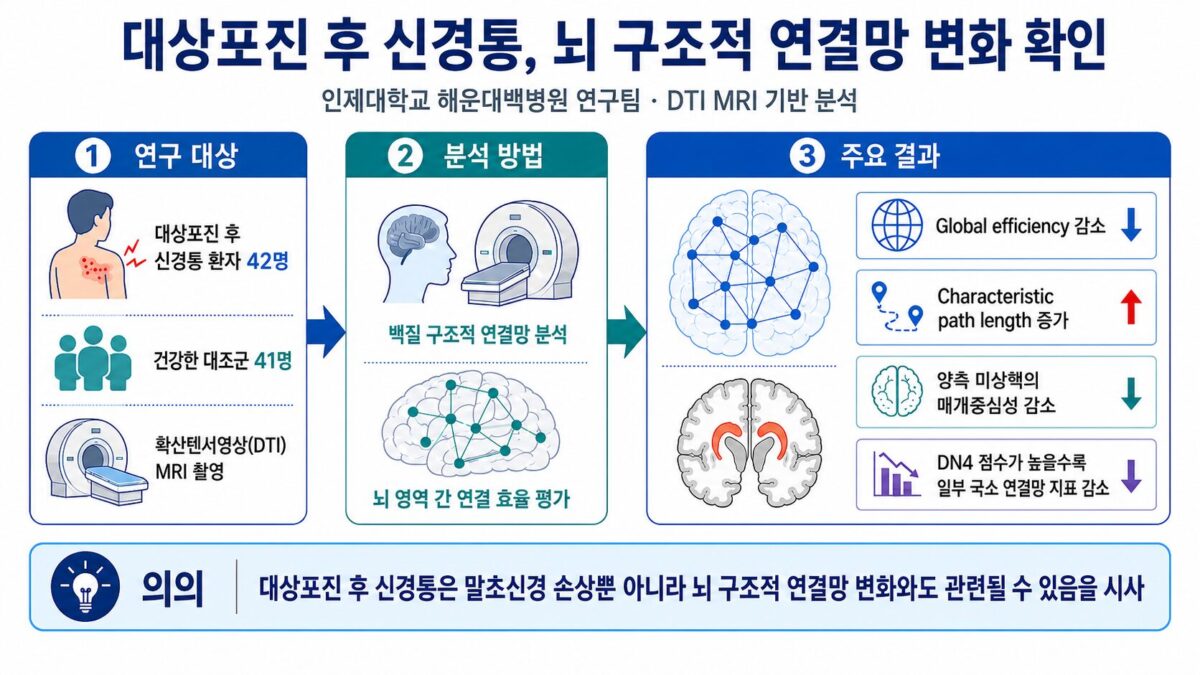
A pioneering research team led by Professor Oh Dae-seok of the Department of Anesthesiology and Pain Medicine and Professor Park Kang-min of the Department of Neurology at Inje University’s Haeundae Paik Hospital has uncovered why. Using advanced brain MRI analysis, the team confirmed that patients suffering from this condition exhibit distinct, abnormal changes in the structural connectivity networks of their brains compared to healthy individuals.
Their findings suggest that the source of this agonizing chronic pain—which can be so intense that even the lightest brush of clothing makes a patient scream—may lie not in the skin or the peripheral nerves, but in fundamental, structural changes within the brain's internal communication pathways.
Shifting the Paradigm from Skin to Brain
For decades, clinicians have generally viewed postherpetic neuralgia as a localized problem resulting from damage to the skin and peripheral nerves during the initial shingles flare-up. However, focusing solely on peripheral nerve injury failed to explain why, after recovering from the same viral infection, only certain patients go on to develop lifelong pain, or why pain patterns vary so widely from person to person.
Suspecting that chronic pain might actually reshape the brain's internal circuitry, the Haeundae Paik Hospital research team turned their focus inward. The team conducted diffusion tensor imaging (DTI) MRI scans on 42 patients diagnosed with postherpetic neuralgia and 41 healthy adults of similar age and sex. DTI is an advanced neuroimaging technique that allows researchers to analyze, at the cellular level, the directionality and structural integrity of the brain's white-matter nerve fibers.
A Breakdown in the Brain's Pain-Control Efficiency
The imaging analysis revealed a striking divergence: patients with postherpetic neuralgia showed a sharp drop in overall brain information-transfer efficiency compared to their healthy counterparts. The neural pathways that act as information highways between different brain regions appeared tangled and lengthened, significantly slowing down the brain's ability to process and suppress pain signals.
Most notably, network relay indicators were markedly diminished in the bilateral caudate nucleus—a critical subcortical structure that serves as the brain's main control center for regulating and suppressing pain. This finding points directly to structural damage within the brain’s built-in pain-control system. Furthermore, the researchers observed a clear correlation: the more severe the patient's self-reported pain, the more distorted their brain connectivity network appeared on the scans.
An Objective Biomarker for Chronic Pain
"Postherpetic neuralgia is a devastating chronic condition that brutally destroys a patient's quality of life," said Professor Oh Dae-seok on July 14. "This study provides strong, objective evidence that this pain extends far beyond simple peripheral nerve damage, and may actually be classified as a chronic brain disease driven by physical changes in the brain's structural networks."
Professor Park Kang-min agreed, emphasizing the long-term clinical significance of the research: "While these findings won’t immediately change how we diagnose or treat patients tomorrow, it is a major scientific breakthrough. We have identified a brain-based biomarker that can objectively diagnose chronic neuralgia. Moving forward, we plan to conduct long-term follow-up studies to develop highly customized treatment strategies and an objective pain-prediction system."
The study’s findings were published in the latest issue (2026, Vol. 39, No. 3) of the prestigious international neuroimaging journal, Brain Topography.