
As the eyes age, gradual changes in the gel-like vitreous humor can trigger floaters—benign black dots, threads, or cobweb-like shapes that drift across the field of vision. When these floaters are mild and do not disrupt daily life, ophthalmologists typically recommend monitoring the condition rather than performing surgery. Over time, the brain naturally adapts to these visual artifacts, making them feel far less bothersome.
However, if these drifting spots are suddenly accompanied by photopsia—flickering flashes of light—or a localized shadow that blocks part of your visual field, a comprehensive eye exam becomes an immediate necessity. Rather than routine, age-related wear, these symptoms are classic indicators of retinal detachment.
The retina is a thin, light-sensitive layer of nerve tissue lining the back of the eye. It detects incoming light, converts it into electrical signals, and transmits them to the brain via the optic nerve to produce the images we see. If the retina separates from its underlying supportive tissue, it loses its blood and oxygen supply. Without timely intervention, this separation can lead to permanent, irreversible vision loss or total blindness.
The Mechanics of Retinal Tears and the Myopia Risk
Retinal detachment most commonly occurs as the vitreous humor inside the eye gradually liquefies and contracts with age. As this jelly-like substance shrinks, it can pull away from the retina. If the vitreous tugs too forcefully, it can tear a hole in the delicate retinal tissue. Liquid vitreous can then seep through the tear, pooling beneath the retina and peeling it away from the back of the eye like wallpaper dampening and falling off a wall.
While historically viewed as an age-related condition, retinal detachment is increasingly threatening younger populations. Adolescents and young adults with severe nearsightedness (high myopia) are particularly vulnerable and require regular screening. In an interview with CoMedi.com, Choi Kyung-sik, a professor of ophthalmology at Soonchunhyang University Seoul Hospital and the legislative director for the Korean Ophthalmological Society, highlighted this regional trend.
"Because South Korea has an exceptionally high prevalence of nearsightedness, the incidence of retinal detachment tends to be higher among teenagers and individuals in their 20s compared to other countries," Choi Kyung-sik explained. "As myopia progresses, the axial length—the front-to-back length of the eyeball—stretches and grows. This physical elongation leaves the retina significantly thinner and much more susceptible to developing tears or breaks."
Beyond age and myopia, other clinical factors can trigger retinal detachment. Inflammatory eye conditions like uveitis can cause fluid accumulation beneath the retina, while advanced diabetic retinopathy can create tractional pulling. If a retinal tear is diagnosed early, it can often be repaired with a non-invasive, in-office laser procedure. However, if the detachment has already progressed extensively, major surgical interventions such as a vitrectomy or scleral buckling become necessary to physically reattach the tissue.
The Danger of Silent Progression to the Macula
In its early stages, a localized shadow or "curtain" in the peripheral vision can be remarkably easy to overlook. This is especially true if only one eye is affected, as the healthy eye will naturally compensate for the deficit, masking the underlying vision loss during daily activities.
The danger escalates dramatically if the detachment spreads to the macula—the highly sensitive central area of the retina responsible for sharp, straight-ahead vision. Once the macula is compromised, central vision drops off precipitously, leaving patients suddenly unable to read, recognize faces, or see clearly.
"If the detachment reaches the macula and affects central vision, sight may not fully recover even after a highly successful surgery," Choi Kyung-sik warned. "Therefore, if you experience a sudden onset of floaters, flashing lights, or a blocked field of vision, you must see an ophthalmologist immediately."
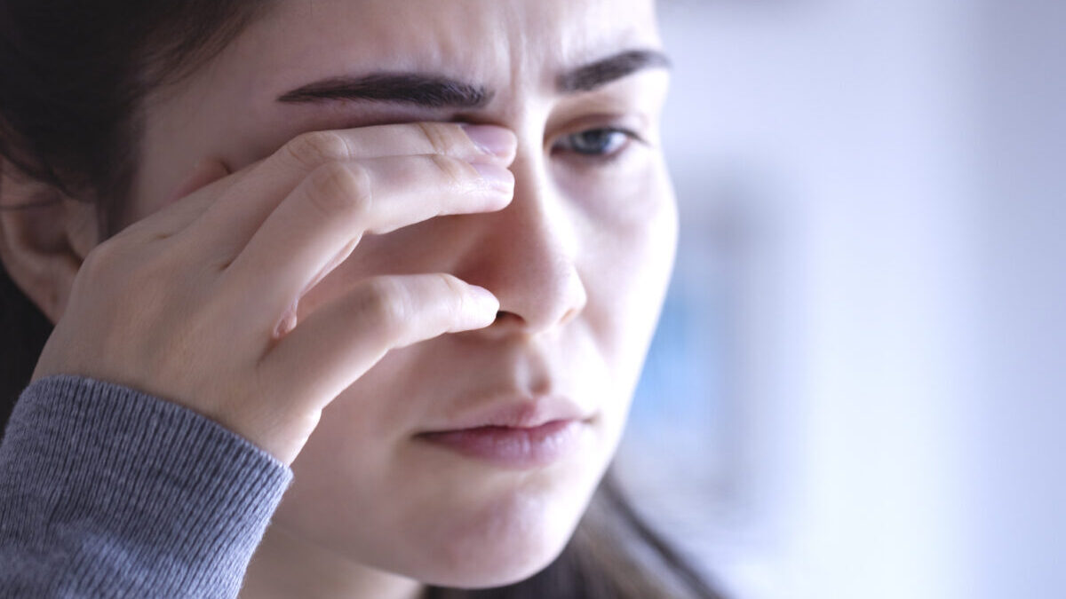
Preventing retinal detachment relies heavily on routine comprehensive eye exams and protecting the eyes from physical trauma. In daily life, this includes avoiding vigorous eye-rubbing. This precaution is particularly vital for patients with atopic dermatitis, who may vigorously rub their eyes to relieve severe itching, unknowingly placing immense physical stress on the retina and risking a traumatic tear.