

Waking up with an unusually intense, pounding headache that offers little relief through over-the-counter painkillers and recurs consistently each morning may be more than mere fatigue or stress. A newly developing headache that is worst upon waking can be a critical warning sign of an underlying brain tumor.
In South Korea, the number of patients treated for brain tumors has climbed steadily. According to National Statistics on Diseases of Public Interest from the Health Insurance Review & Assessment Service, the number of individuals treated for benign brain tumors rose 28% to 66,350 in 2025, up from 51,842 in 2021. Meanwhile, malignant brain tumor cases increased 13% over the same period, rising from 11,945 to 13,488.
Brain tumors can disrupt a wide array of neurological functions—including movement, speech, sensation, and memory—depending on their precise anatomical location. Even non-malignant tumors can cause severe neurological issues as they grow and compress adjacent brain tissue and nerves.
Headache is the single most common symptom. Medical specialists urge caution when a patient experiences a novel headache—one distinctly different from past episodes—that is most severe in the morning and progressively worsens over time. Additional symptoms vary by tumor site and can include visual field defects, persistent nausea and vomiting, sensory abnormalities, speech impairment, and seizures. Tumors located in the frontal or temporal lobes can also impair memory and make emotional regulation increasingly difficult.
Because these symptoms mimic other conditions, patients often seek care in the wrong medical specialties. Many initially consult ophthalmologists for vision changes, gastroenterologists for unexplained vomiting, or psychiatrists for sudden behavioral and emotional shifts. Navigating between multiple clinical departments frequently leads to delayed diagnoses.
Treatment strategies are tailored to the tumor’s type, location, and size, as well as the patient's overall health, incorporating surgery, radiation therapy, and chemotherapy either individually or in combination. The primary goal of surgical intervention is to achieve maximum tumor removal while preserving normal brain tissue and function.
Prognosis varies significantly depending on whether the tumor is benign or malignant. According to the National Cancer Information Center, five-year survival rates for common benign brain tumors remain exceptionally high: meningiomas at 95%, pituitary adenomas at 97%, and schwannomas at 94%. Conversely, the five-year relative survival rate for malignant brain and central nervous system cancers stood at 39.1% between 2019 and 2023, underscoring the vital importance of early detection.

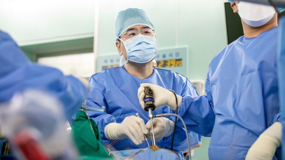
Medical advances have expanded treatment options, particularly through minimally invasive techniques. "Minimally invasive approaches are primarily used for skull base lesions such as pituitary tumors," said Seo Young-beom, a professor of neurosurgery in the Brain Tumor Division at Korea University Ansan Hospital. "By inserting an endoscope and specialized instruments through the nasal cavity, we can access the lesion without performing a traditional craniotomy, thereby minimizing unnecessary damage to healthy brain tissue and enabling faster patient recovery."
When surgical intervention carries a high risk of nerve damage or is anatomically difficult, stereotactic radiosurgery offers a viable alternative. After precisely mapping the lesion using MRI and CT imaging, this non-invasive technique delivers targeted, high-dose radiation directly to the tumor. The procedure is painless, requires no skull incision, and significantly reduces physical strain on the patient.
Because the exact causes of brain tumors remain largely unknown, definitive preventive measures do not yet exist.
"If symptoms such as persistent headaches or visual field impairments fail to improve with standard treatment, patients should consider whether the underlying cause originates in the brain," Seo said. "In such cases, visiting a neurosurgery department for specialist evaluation and undergoing detailed diagnostic imaging, such as an MRI or CT scan, is essential for securing a timely diagnosis and starting effective treatment."