

South Korean researchers have developed a breakthrough MRI technique that images the body’s lymphatic system without an invasive groin injection. The new method relies instead on a standard intravenous line in an arm vein to produce remarkably clear images of lymph vessels deep within the liver and mesentery—critical anatomical regions that have historically been nearly impossible to evaluate with existing diagnostic tests.
A research team led by Prof. Heo Se-beom of the Department of Radiology at Seoul National University Hospital, Prof. Yoon Sung-hwan of the Biomedical Research Institute, and Prof. Kwon Ryeo-min of the Department of Radiology at Hallym University Hospital announced on the 9th that they developed this new imaging modality, termed “intravenous contrast-enhanced MR lymphangiography (IV-MRL),” and successfully verified its clinical efficacy. Their findings were published in the latest issue of Radiology, the leading international journal in the field.
The lymphatic system serves as a vital biological network maintaining systemic immunity and fluid balance. When these delicate pathways are compromised during major interventions like cancer surgery, lymphatic fluid can leak into internal cavities. This leakage triggers severe complications, including acute malnutrition, weakened immune defenses, and chylous ascites—the dangerous accumulation of milky, fat-rich lymph fluid inside the abdomen. While it sounds uncommon, clinical data reveals that such leaks occur in approximately 6 to 7 out of every 100 patients following colorectal cancer surgery alone.
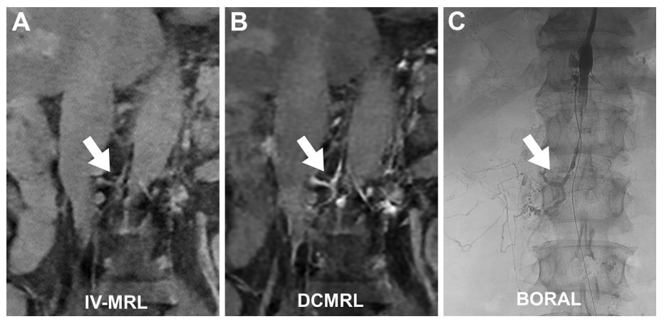
Pinpointing the exact site of a lymphatic leak, however, has remained an operational nightmare for clinicians. Standard diagnostic procedures—namely intranodal lymphangiography and dynamic contrast-enhanced MR lymphangiography (DCMRL)—require radiologists to precisely guide a needle directly into a groin lymph node. This approach is highly examiner-dependent, technically demanding, and routinely fails to visualize the liver and mesenteric lymphatics, which accounts for more than 70% of the body's total lymph production. Past domestic studies show that the success rate of this groin-dependent approach fluctuates heavily based on the patient's underlying condition, dropping from 94% down to 82%. For patients suffering from chylous ascites, the localization success rate plummets to a mere 55%.
Visualizing the Lymphatic Phase
To overcome these diagnostic blind spots, the research team initially administered the intravenous contrast dye to animal models, tracking the concentration dynamics between the bloodstream and lymph fluid over a timeline. They discovered a distinct, predictable “lymphatic phase” where the contrast concentration within the lymph fluid surpassed that of the surrounding blood supply, beginning 20 to 30 minutes post-injection. On MRI scans captured during this optimal window, the signal intensity of the cisterna chyli—the crucial lymphatic reservoir located just below the thoracic duct—surged to 5.5 times that of the paraspinal muscles, bringing the complex web of lymph vessels into sharp relief.
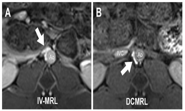
The researchers subsequently moved to a pilot clinical study tracking 16 patients (mean age 56.6 years) suspected of suffering from active lymph leaks between September 2024 and August 2025. The clinical trials mirrored the animal models precisely. Just 1 minute after the arm injection, the lymph-vessel-to-vein signal ratio stood at a faint 0.49. However, by the 20-minute mark, the ratio climbed significantly to 1.47, providing a stark visual contrast. While conventional groin-injection methods could only map out the thoracic duct and retroperitoneal tracks, the non-invasive intravenous method successfully illuminated the liver and mesenteric vessels in full detail.

“Unlike conventional lymph-node injections, IV-MRL utilizes a standard, peripheral IV line, which dramatically lowers the technical difficulty of the procedure and spares the patient from a painful, invasive groin puncture,” Prof. Heo Se-beom explained. “We expect this technique to significantly elevate diagnostic accuracy for complex lymph-leak disorders and streamline the formulation of targeted treatment strategies moving forward.”
Until now, even top-tier medical institutions globally were forced to insert needles directly into internal organs to obtain clear images of the liver and mesenteric lymphatics. By unlocking full-body lymphatic mapping through a simple, single injection in the arm, this innovative approach provides a definitive roadmap to locate hidden fluid leaks anywhere in the body entirely needle-free.