
When surgeons reattach a torn shoulder tendon to the bone, a frustrating setback often occurs over time: the repair tears again. The root cause is rarely a failure of the surgical sutures themselves. Instead, the body struggles to regenerate the complex natural transition between tendon and bone, filling the gap with structurally weak scar tissue.
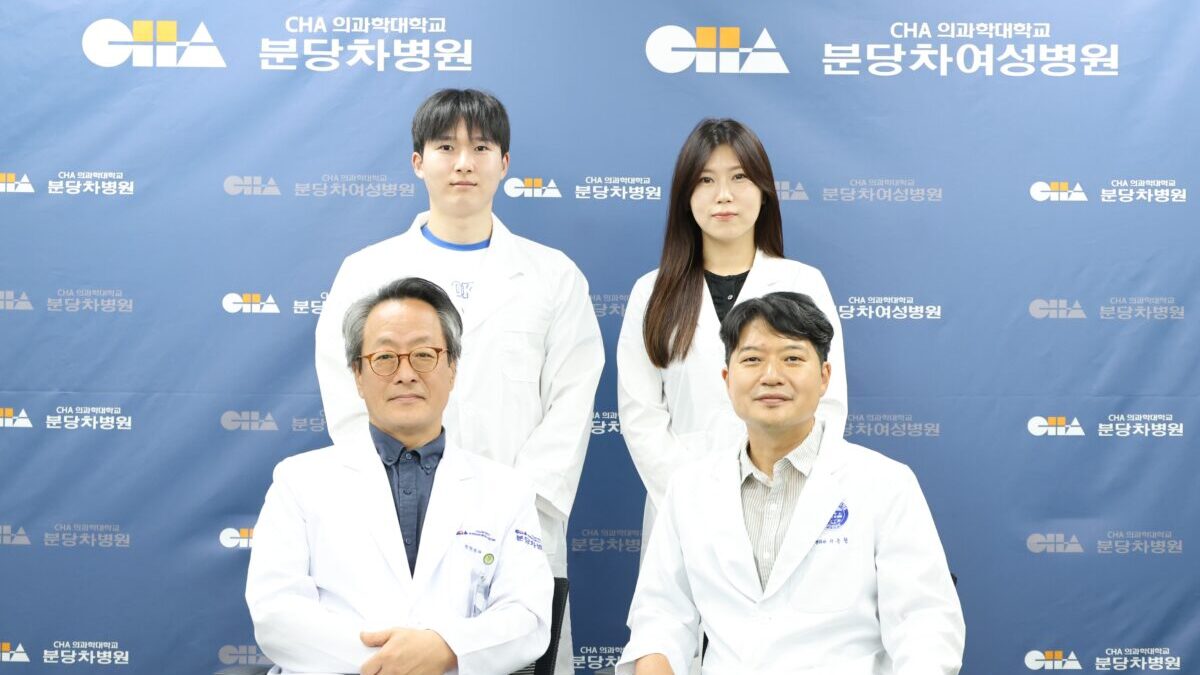
To overcome this fundamental limitation, a research team at CHA University’s Bundang CHA Hospital developed a tissue-engineering strategy designed to reconstruct the natural tendon-to-bone junction. Led by orthopedic surgery professors Kim Jae-hwa and Lee Soon-chul, the team created a single "dual-gradient" artificial scaffold paired with an "exosome mimetic" to guide step-by-step tissue repair. Their findings were published in the bio-materials journal Small and featured on the Biological Research Information Center (BRIC) list of "People Who Made Korea Shine" (Hanbitsa).
The Enthesis Challenge: Why Repaired Tendons Fail
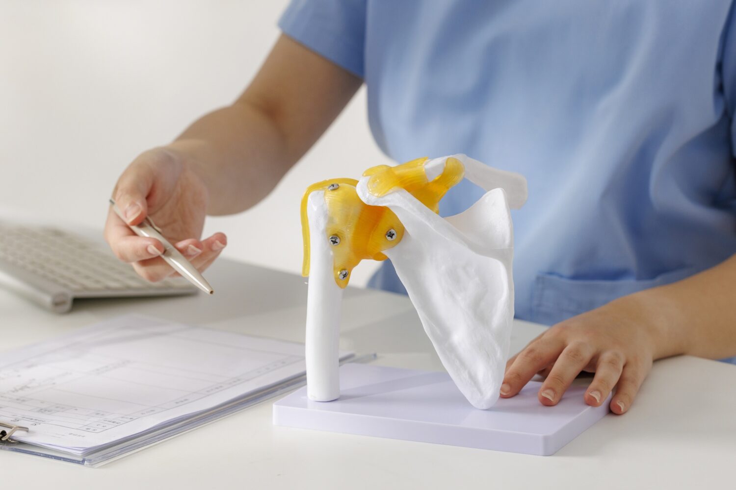
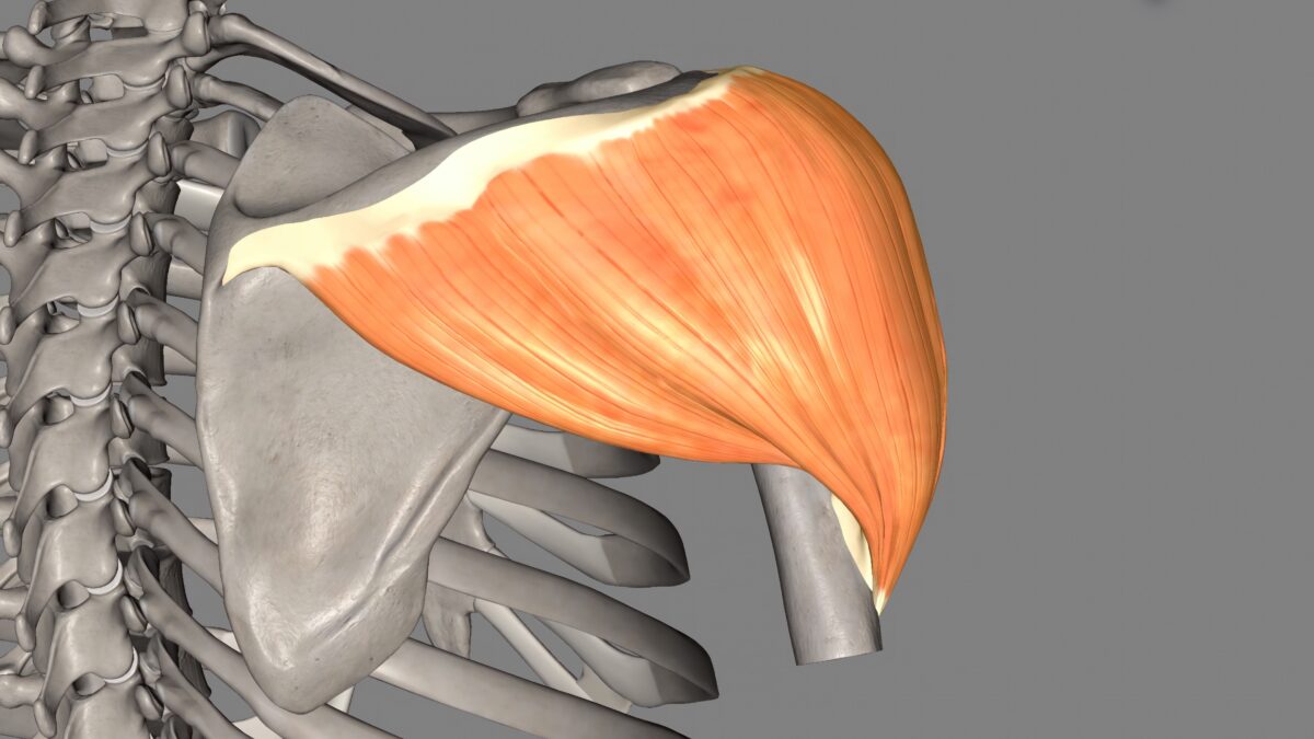
The rotator cuff is a group of muscles and tendons surrounding the shoulder joint that enables arm lifting and rotation. Degenerative age-related changes or repetitive overhead strain can cause these tendons to tear. When pain is severe or functional loss is substantial, surgeons perform an operation to re-anchor the torn tendon to the head of the humerus.
However, a healthy tendon-to-bone junction (enthesis) is not a simple joint where soft tissue directly meets hard bone. It features a continuous, multiphasic gradient that transitions gradually from tendon to uncalcified fibrocartilage, calcified fibrocartilage, and finally bone. This specialized architecture dissipates the mechanical stress generated during shoulder movement. Following standard surgery, the body fails to recreate this gradient, depositing disorganized fibrous scar tissue that lacks original structural integrity.
Consequently, post-operative re-tear rates remain notably high. A meta-analysis of 31 studies found overall re-tear rates between 15% and 21%. For large and massive tears, the re-tear rate reached 37%, with advanced age and severe muscle fatty infiltration further elevating the risk. Because healing remains delicate, non-surgical management like physical therapy is often preferred for smaller tears, while surgical patients require carefully tailored post-operative rehabilitation rather than rushed mobilization.
A Dual-Gradient Scaffold with Staged Molecular Delivery
To replicate the natural enthesis, the research team engineered a single scaffold rather than layering separate materials—an older technique that frequently suffered from layer separation. In this unified dual-gradient scaffold, both fiber alignment and mineral concentration shift progressively from one side to the other. The tendon side supports tenocyte proliferation, while the bone-contacting side induces fibrocartilage and bone formation.
The scaffold works in tandem with an "exosome mimetic" designed to imitate extracellular vesicles involved in cellular communication. The system delivers therapeutic signals in sequence corresponding to each healing phase: shortly after surgery, it suppresses local inflammation and oxidative stress; later, it releases regenerative substances that encourage tendon and bone growth. The biodegradable matrix acts as a temporary physical platform for cell growth before safely breaking down inside the body.
Pre-Clinical Success and Future Clinical Outlook
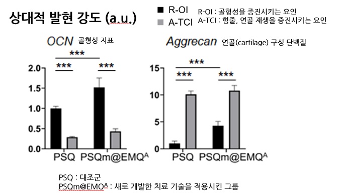
In animal models of rotator cuff tears, combining the dual-gradient scaffold with the exosome mimetic produced marked structural improvements over conventional repair techniques. Treated animals exhibited enhanced fibrocartilage and bone formation, tighter fiber alignment resembling native tissue, and reduced fatty degeneration. Biomechanical testing demonstrated that the regenerated junction withstood significantly higher tensile force, confirming a firmer physical connection.
"This study moves beyond simple mechanical suturing to offer a new therapeutic strategy for regenerating the tendon-to-bone interface into tissue that closely resembles native anatomy," said Professor Kim Jae-hwa. "We anticipate this technology will eventually extend to various orthopedic procedures where tendons or ligaments anchor to bone, including Achilles tendon and anterior cruciate ligament repairs."
While human clinical trials are still required to verify safety and confirm whether the technique reduces re-tear rates in clinical practice, the approach represents a promising step toward long-term joint restoration.