
Eye photos taken during routine health screenings could help identify who currently has dementia and predict who is at risk of developing the neurodegenerative condition years later, according to new research from Seoul National University Hospital.
A joint research team led by Prof. Park Sang-min of the Department of Family Medicine, Dr. Jang Ju-young of XaiMed Co., Research Assistant Prof. Han Chang-ho of the Seoul National University College of Medicine’s Medical Big Data Research Center, and Dr. Kim Jae-won of the Department of Biomedical Sciences developed the deep-learning system after analyzing 108,008 retinal fundus photographs from 36,322 health-screening participants.
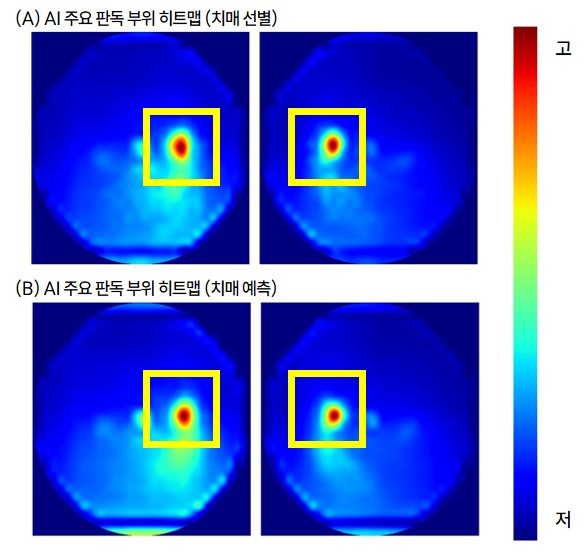
The team evaluated not only the algorithm’s predictive accuracy but also the visual features it relied on. In assessing both current cognitive status and future disease risk, the AI consistently focused on the optic disc—the convergence point of the optic nerve—and its surrounding regions.
Connecting the Retina to the Brain
A retinal fundus photograph is an image taken through the pupil that captures the retina, optic nerve, and blood vessels inside the eye. It is routinely performed during eye examinations and general health checkups.
Researchers turned to the eye because the retina functions as an extension of the central nervous system. Subtle neural or microvascular changes in the brain often reflect in the eye, offering a non-invasive window into neurological health.
Drawing on health-screening data collected at Seoul National University Hospital between 2004 and 2016, the team identified 1,001 individuals who were diagnosed with dementia and prescribed related medication within two years before or after imaging (comprising 2,868 fundus photos). They trained the model by comparing these images against a control group without dementia. Among multiple model architectures, a deep-learning network pre-trained on a large volume of fundus images yielded the highest performance.
Outperforming Traditional Clinical Risk Scores
The AI system achieved an area under the curve (AUC) score of 0.750 in distinguishing individuals with current dementia from healthy controls (where a value closer to 0.5 indicates performance no better than chance, and 1.0 represents perfect discrimination).
To benchmark the tool, researchers compared its performance against the Cardiovascular Risk Factors, Aging, and Dementia (CAIDE) score, a widely used clinical tool that estimates long-term dementia risk based on age, sex, education level, blood pressure, body mass index, cholesterol, and physical activity without requiring brain imaging. While CAIDE scored 0.624 for identifying current dementia, the fundus-photo AI scored 0.750.
The gap was even wider in predicting future dementia risk: the AI achieved an AUC of 0.812 compared to CAIDE’s 0.689. The AI’s predictive advantage persisted even after controlling for conventional demographic and cardiovascular risk factors, indicating that retinal images capture unique neurological biomarkers that traditional assessments miss.
The Optic Disc as a Primary Decision Point
To understand the AI's decision-making process, the team mapped the specific visual regions the model prioritized. The primary focal point was the optic disc—the exit site where nerve fibers carrying visual information gather to enter the brain.
The model’s attention to the optic disc was 5.72 times higher than to adjacent background areas. High attention was also concentrated along the nerve fiber pathways extending toward the macula. Because prior neuroimaging studies have linked structural changes in these specific retinal areas to cognitive decline, the findings confirm that the AI responded to authentic biological markers rather than artifacts in the images.
Opportunistic Screening in Primary Care
The findings suggest that existing fundus photos taken during standard health checkups could be leveraged for opportunistic dementia screening without requiring additional, expensive imaging.
If validated further, the technology could serve as an early triage tool in primary care settings, identifying high-risk individuals and referring them for comprehensive cognitive testing or specialist care. The researchers stressed, however, that the AI is not a standalone diagnostic tool, as a formal dementia diagnosis still requires clinical evaluation, cognitive assessments, and, where necessary, brain MRI or fluid biomarker tests.
Because the study relied on retrospective data from a single medical center, additional validation is required across diverse healthcare settings, age groups, and racial backgrounds.
"We hope this AI technology, which utilizes routine screening data without high-cost diagnostic procedures, will become a valuable decision-support tool for dementia prevention and early intervention strategies," said Prof. Park Sang-min.
Dr. Jang Ju-young added, "By evaluating not only predictive performance but also model explainability and clinical utility, this study establishes an important direction for developing fundus-image-based AI biomarkers."
The study was published online July 4 in the journal npj Digital Medicine.